Karly Dawson, PA-C, Brian Gilmer, MD
Introduction
The peroneus longus and brevis are muscles which begin high on the outer aspect of the lower leg (near the knee) and become tendons as they approach the ankle. Together they serve to move the foot inward and outward and help stabilize the ankle joint.
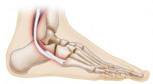
Anatomy
Peroneus longus and peroneus brevis are muscles that originate on the outer bone of the lower leg called the fibula. These become tendons above the ankle in order to attach the muscles to the foot. The two tendons sit in a groove behind the distal fibula and are held in place by fascia and ligamentous tissue called the peroneal sheath and sling respectively. The peroneus brevis tendon is located directly behind the fibula bone and in general is more prone to injury. It serves to evert the foot, meaning to move it outwardly away from the rest of the leg. The peroneus longus sits behind the brevis tendon and wraps underneath the foot to assist in eversion and also to flex the foot downwards.
Injury
Peroneal Tendonitis is a condition where one or both of the peroneus tendons become enlarged and inflamed. This is generally an overuse injury that can be due to repetitive motions. It can also result from a rapid increase in activity or an acute trauma like an ankle sprain.
Peroneal Tendinosis is a chronic condition that refers to inflammation and tearing of one or both of the tendons that occurs over a longer period of time. The peroneus brevis is more commonly affected. This can be due to having multiple traumas such as sustaining repetitive ankle sprains, or from repetitive subluxations, which is where the tendons slip out of their normal groove behind the fibula. The tendons fray and stretch as they are rubbed against the edge of the fibula and this causes degenerative tearing.
Tendon subluxation or dislocation usually occurs as the result of an associated ligamentous injury. The peroneal retinaculum holds the ligaments in place beneath the fibula, but injury to this structure can allow them to slip out of their normal position and lead to tearing of the tendons and dysfunction.
The lateral ankle ligaments (anterior talofibular ligament(ATFL)andcalcaneofibularligaments (CFL)) are located just below and in front of the tip of the fibula. Injury to these structures is commonly referred to as an ankle sprain. If these are injured or torn, the resultant instability can lead to peroneus tendon injuries as the muscles attempt to stabilize the ankle and compensate for the ligamentous insufficiency.
Symptoms
Both acute and chronic peroneal tendon injuries cause lateral (the side farthest from the other foot) ankle pain and swelling. Acute injuries can also cause redness or bruising and weakness, while chronic injuries can additionally lead to catching or a sense of instability.
Diagnosis
Physical examination can identify certain related conditions, such as laxity of the ankle ligaments or a dislocated tendon. X-rays may be taken to rule out fractures or other bony abnormalities. An MRI may ultimately be obtained to evaluate the integrity of the peroneus tendons and their surrounding ligaments.
Treatment
The severity and chronicity of these injuries determines the treatment course. Inflammatory and overuse type injuries are usually responsive to conservative management, though it may take several weeks to months to notice full resolution of symptoms. Dislocations of the peroneus tendons or acute tears will often require more prompt surgical intervention.
Non-operative Management
First line treatment of overuse type injuries include rest, ice, elevation, anti-inflammatories, and activity modification. We may recommend a course of physical therapy to maintain range of motion along with guidance in strength and balancing exercises. If there is a dislocation or an acute peroneus tendon tear, chronic lateral ankle instability, or severe interruption of a patient’s life from a peroneal tendon injury despite an adequate rehabilitation program, surgical options may need to be considered.
Operative Management
If a peroneus tendon tear is identified, we may recommend “tubularization” to repair it. This method includes sewing up the torn area of the tendon to restore it to its normal shape and smoothness. Sometimes a groove deepening procedure of the distal fibula is also required to help provide a more stable location for the tendons to remain in place if instability of the tendons is present. Peroneal tendon repair surgery may be accompanied by a lateral ankle stabilization method (a Brostrom procedure for example), which repairs and reinforces the torn ligaments in order to create a more normal environment for the peroneal tendons and prevent further injury.
Recovery
After surgery patients are instructed to remain non-weight bearing on the operative leg for a period of approximately 6 weeks after the surgery to allow for healing and to prevent re-injuries. Patients are enrolled in physical therapy during that time and will continue with therapy once weight bearing is permitted to restore strength and help prepare them for sport-specific demands. Total recovery time after surgery varies but may take up to 3-6 months to return to full high impact activities.
For more information about our services or to schedule an appointment, call us at / / or click here to request an appointment online. We’ll respond to you as soon as possible.