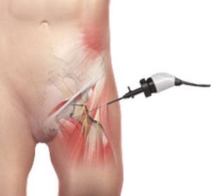
Hip arthroscopy is one of the most rapidly developing fields in orthopedic surgery. It is a minimally invasive surgery to correct problems in and around the hip joint. This surgery involves making 2 or 3 tiny “poke” holes in the front of the hip and placing an arthroscope, a special camera that allows one to look inside a joint, through one of those holes. The other one or two small holes are used to place special instruments inside the joint to perform the surgery.
Unlike the shoulder and the knee, the hip is a very tight ball-in-socket joint. Getting into the joint can be challenging. However, there are two things that allow surgeons to get into the hip. The first and most important is the use of a special operating room table which allows the surgeon to gently pull on the patient’s foot to distract, or open up, the hip joint. An x-ray machine allows the surgeon to see the amount of distraction before any incision is made. A special well-padded post is placed against the patient’s perineum to pull against. Also, an arthroscopic pump is used to push sterile water into the joint and distend, or expand, the joint. This provides a space for surgeons to see and work.
Indication for surgery
The indications for hip arthroscopy, or the reasons this surgery is performed, have expanded over the last decade as equipment has improved, and new techniques have evolved. The most common reason is a condition called femoroacetabular impingement, or FAI. As stated, the hip joint is a very tight ball-in-socket joint. In FAI, there is a physical mismatch between the shape of the ball and socket, like a square peg in a round hole. Imagine if the ball is not exactly round, or if the rim of the socket has a slight boney overhang. In certain positions, the two shapes don’t match which leads to trauma to the cartilage in the joint or leads to a tear of a structure called the labrum. The labrum is fibrocartilaginous ring which essentially surrounds the socket. Because of this shape mismatch, FAI leads to arthritis. In fact, FAI is the most common cause of arthritis in the hip. It is hoped that by correcting this shape mismatch early, surgeons can prevent hip arthritis. This has led to a new field in orthopedics called hip preservation. Whether arthritis is prevented or not is unclear at this time because FAI is a relatively new diagnosis with no long term data. However, the short term data has shown 80% good-to-excellent outcomes in terms of patient satisfaction and hip function. One study specifically involving intercollegiate athletes showed 90% of patients returned to sports after FAI surgery.
The other indications for hip arthroscopy include labral tears without FAI, loose bodies in the joint, hip instability, tears of a structure called the ligamentum teres, an entity called snapping hip syndrome, and abductor tendon tears. Hip instability, even if subtle and in the absence of trauma, can be quite painful and cause significant dysfunction. The ligamentum teres is a small ligament in the hip joint which connects, or tethers, the ball to the socket. This structure has been torn in seemingly insignificant trauma and is also a source of pain and disability. The painful, snapping hip can be caused by two different structures, the iliopsoas and the iliotibial band, which, while usually treated successfully non-operatively, can lead to surgical treatment. The hip abductors, specifically the gluteus medius and minimus, are two muscles that attach to the outside of the hip and can tear. They sometimes need a repair similar to the shoulder’s rotator cuff.
Contraindication for surgery
The contraindications for hip arthroscopy include severe arthritis of the hip and avascular necrosis, or other conditions of the back or lower extremities that make distracting the hip joint unsafe or impossible. Relative contra-indications include confounding pain from other sources (i.e., the lumbar spine), failure to improve with a diagnostic hip injection, and not having tried non-operative treatment.
Recovery
Recovery from hip arthroscopy is faster than with open surgery and is less painful. In fact, most patients are off narcotic pain medication within 3 days of surgery. The surgery is performed in an outpatient status with the patient going home after surgery either the same day or the next morning. Patients are usually on crutches for the first three weeks and in some cases wear a brace for the first six weeks. At home, patients are encouraged to start using a stationary bike as soon as possible after the surgery to begin restoring range of motion. There is physical therapy once or twice a week for the first few months after surgery to work on range of motion followed by strengthening. Most patients are able to return to sport in 4 to 6 months.
Complications
As with all surgical procedures, complications may occur with hip arthroscopy. Fortunately, these are very uncommon. The most common complication involves injuring the cartilage or labrum attempting to gain access to the hip joint. This complication can be minimized with experience and by carefully following a strict technique to enter the joint. Further, there has been no study to demonstrate that these iatrogenic chondrolabral injuries are of any long term consequence. The next most common complication is a stretch or compression nerve injury (usually resulting in groin and/or genital numbness) which resolves in 99% of the cases within the first 6 weeks. Other complications include heterotopic bone formation and the formation of a blood clot in the leg called a deep venous thrombosis, or DVT. Heterotopic bone formation occurs when the body forms bone in the soft tissues after a trauma or a surgery for unknown reasons. This bone often causes no problems, but sometimes may cause significant stiffness. Patients should take naproxen for three weeks after surgery to help prevent this bone formation.
Conservative vs. surgical
Treating the hip using conservative measures offers us time to see if we can strengthening the surrounding tissue and provide relief with an ultrasound guided hip intra-articular injection. The injection has both therapeutic and diagnostic benefits. Combining the injection with a dedicated physical therapy program, you may get enough relief to prolong the need or eliminate the need of surgery. If conservative treatment fails and you are indicated for a hip arthroscopy we would then discuss the details of surgical intervention.
Preparing for your hip arthroscopy
Physical therapy: You should have a short visit with physical therapy prior to your surgery to learn the proper post op mechanics and weight-bearing precautions. Also, if you are doing your physical therapy by someone other than the Mammoth Sport Center, you will need to make sure your first physical therapy visit is scheduled within 3-5 days of surgery. If you are doing therapy with Mammoth Sport, you should receive a phone call about 2 weeks ahead of your surgery date to schedule your therapy. If you don’t hear from them, please call to schedule an appointment. If your surgery is on a Wednesday, either Friday or Monday is appropriate for your first appointment.
Crutches: Crutches will be utilized for approximately the first 3 weeks while you will need to properly monitor the amount of weight you are placing on your foot. It is important that you are using a flat foot walking technique to minimize any continuous use of the muscles on the front of your hip. If you are in a toe-touch stance or non-weightbearing, you can increase your chance of overusing the muscles and creating a secondary overuse injury.
MRI: An MRI with a special advanced protocol may be needed pre-operatively to view the integrity of the labrum and cartilage. Your surgeon will discuss this with you ahead of time.
CT 3D Reconstruction: A CT 3D reconstruction will provide a 3D model of the bones of your hip which will allow your surgeon to view the shape of your femoral head and femoral neck. By getting this diagnostic study, your surgeon will be able to map out what needs to be removed so reshape your bone to make it anatomically correct.
Bracing: In a minority of cases this may be used after surgery. You will be fit for a brace before surgery if appropriate.
Work/school: Ask about a work or school note before surgery if you need one. Generally students and those with sedentary or desk-type jobs can go back to school or work the Monday following surgery. This is much more variable for those with labor intensive jobs, and can depend on the specifics of your job duties, so please ask ahead of time.
Scheduling your surgery
Our surgery coordinators, Leticia Bravo and Heather Tindall, RN, will help to schedule your surgery at Mammoth Hospital. They will contact you for a telephone interview and review your health history. If it has been some time since your visit with Dr. Crall, you will also need to visit with Bart White, PA-C, his physician’s assistant, closer to the time of surgery.
What to expect on your surgery day
Typically you will be asked to arrive at your surgical location 1.5 hours prior to your anticipated surgery time. You will be called in the late afternoon on the day prior to your operation day and told the time. This will allow the pre-operative nurses to do their evaluation, start your IV and allow you to complete any remaining paperwork. This will also give you the opportunity to meet your anesthesiologist.
It is important not to eat or drink after 12 midnight on the night prior to surgery. This will reduce your risk of vomiting and avoid potentially having your surgery rescheduled. If you are taking medications you may do so with a (small) sip of water however, please review all your medications with the physician assistant at the time of pre-operative visit to make sure they will not interfere with your surgery or anesthesia.
You will need a responsible adult to drive you home when you are discharged from the hospital. It is preferable for that person to be in the surgical facility when your surgery is done so your surgeon can communicate the operative findings. Although every effort is made for the surgeon to discuss these findings directly with you, sometimes the lingering effects of anesthesia can make memories of this a little foggy. We will also send you an electronic report of your surgery, so you can review this at home once you have recovered more from anesthesia.
A patient guide for post-operative recovery and rehabilitation
The following guide is an overview of what patients should expect during the weeks and months following Hip Arthroscopy. This guide may help to answer common questions or concerns that come up after this surgery. Please refer to your surgeon and physical therapist for specific questions and exact guidelines of your recovery.
Day One: You just came out of recovery. At this time, you have not quite gotten back to feeling like yourself. The recovery team will go over your precautions and guidelines with you. Your hip is very fragile, and it is very important to know your “weight-bearing precautions”.
Wound: If your wound is oozing from the surgery site and the dressing site appears soaked with bloody fluid, please change the dressing as needed. This normally occurs from fluid irrigation during surgery and should resolve within 24-36 hours. You may remove the dressing on post-op day #2. Apply band aids to wound sites and change them once a day. Keep the wound clean and dry; please do not use Bacitracin or other ointments. Showering is allowed on post-operative day #4 if the wound and dressings are dry. Just pat the wounds dry with a towel when you are done. Don’t submerge the incisions in a tub or bath for 2-3 days after your sutures are removed in the office.
Medications: The anesthetic drugs used during surgery may cause nausea for the first 24 hours. If nausea is encountered, drink only clear liquids. The only solids should be dry crackers or toast. If nausea and vomiting become severe of the patient shows signs of dehydration (lack of urination), please call the doctor or hospital and ask to speak to the Orthopedic Surgeon on call. Local anesthetics are put into the incision at surgery. It is not uncommon to experience more pain as they wear off on the first or second day after surgery. This is also the time when swelling peaks. Taking pain medication prior to bed will aid in sleeping.
- Heterotopic bone prophylaxis/anti-inflammatory for 10 days: Naproxen (over the counter strength—500mg tablets). One tablet by mouth twice daily (morning and night) x 21 days
- Pain: Percocet 5/325 mg tablets, 1-2 tablets every 4 hours as needed for pain
- If you have pre-existing problems with heartburn/GERD: Stomach ulcer/reflux prophylaxis: Prilosec OTC 20mg by mouth daily
- If you have a history of blood clots/DVT or are at high risk for blood clots, you will be advised to take aspirin or a full strength blood thinner for 3 weeks after the surgery.
- If you have other problems after surgery (intractable nausea, spasms, etc.) let us know and we can provide medications for these problems as well.
Weight-bearing: Typically you are able to put about 20 pounds of pressure on your repaired side. A little weight on your foot actually takes some of the pressure off the repaired hip.
Brace: You will be instructed on this if it is needed prior to the surgery, and again before your discharge home from the hospital.
Weeks 0 – 3: Within the first several days after surgery, you will have your first physical therapy appointment. You may be a little out of it at this point and may be experiencing a significant amount of pain. The first session you should expect to have your bandage removed and a smaller one put in its place. You will also be taught, in more detail, how to use your crutches, brace, and any precautions. The therapist will also help you move your hip through a safe range of motion and start performing very gentle exercises. You will also be given a written protocol, so you will know what to expect. You will be instructed on exercises you can do at home. You will be encouraged to use a stationary bike to help keep the muscles moving. This phase is very important to protect your hip repair. Avoid putting too much weight on your leg and lifting the leg up (using your hip flexor muscles). Your surgeon recommends avoiding repetitive active hip flexion (lifting your leg up at the hip) until 2-3 weeks after your surgery. This precaution is to prevent excessive hip flexor tendinitis after your surgery.
In therapy, you will receive specific stretching and muscle work to the front of your belly (where some of your hip muscles start) and to the front, inner, and back side of the hip complex. You will also start some gentle strength exercises for the muscles around the hip complex. The goals of this stage are to restore the function of the hip, back and leg muscles in order to prepare them for use once you start walking.
Weeks 3 – 5: This is an exciting time. Depending on what the surgeon states, you will usually stop using the crutches and brace. You may need to wean off the crutches, going from using both crutches to using one crutch, and then to none. It’s important at this phase to use the crutch in the opposite arm of your surgery. Contrary to popular belief, using the crutch in the opposite side reduces the stress at the hip. Using the crutch on the same side causes more stress. Your hip should be feeling much better at this point, but be careful to avoid stressing the repaired labrum and hip muscles.
Exercises: You will start gentle hip flexion at this point, but don’t overdo it. You may cause tendinitis at this area if you are too aggressive. Your therapist will start more exercises at this point to strengthen the gluteus muscles (muscles that make up the buttocks), hip inner and outer thigh muscles, and back (core) muscles. These should all be well tolerated and cause little to no stress on the surgery site. You will receive more home based exercises at this point to progress your mobility. Gait training (walker training) will also be performed to help you return to walking smoothly with no limp. You may need work on balance over the newly repaired hip. Balance boards will be used at this point. Bicycling is also encouraged now.
Manual therapy: This will continue to help stretch out your muscles, loosen them up and help with strength training. Work will also progress on your scar sites to make them move more easily. Gentle hip joint stretching may be used early in the recovery with more advanced stretching used later in the recovery.
Aqua Therapy: When your surgical sites are fully healed, you may be encouraged to begin pool therapy for cardiovascular exercise.
Weeks 6 – 9: At this point, the hip should be feeling pretty good. Some stiffness, tightness or soreness may be experienced, especially in the groin area. At this phase, self-stretching becomes more important and you will have home strengthening to do. Your walking should be without a limp, or you should be working on walking smoothly.
Manual Therapy: Your therapist will generally continue to perform deep muscle stretching and add more aggressive joint stretching. This may include using a strap or belt to help pull the socket and restore full functional mobility of the hip. You will most likely have your hip stretched in several different directions to restore the leg’s ability to move well. Mild soreness may be experienced, but sharp pain should not. Full hip range of motion is the goal at this point.
Exercise: You will be advanced with leg and hip strength training. These exercises will include Pilates-type training, closed-chain exercises (leg presses, step training, and balance work), and open-chain exercises (proprioceptive neuromuscular facilitation, or PNF, hip patterns to help work hip flexors which have been avoided until this point). More advanced leg stretching will be prescribed by your therapist to help restore full motion to your hip.
Cardiovascular: Advanced training on the bike will continue at this point.
Weeks 9 – 12: The goals of this stage are to restore full range of motion through stretching, strength training, and “functional” training.
Manual Therapy: As noted above, you will continue to have skilled manual therapy to ensure your hip is moving well. End range stretching will be advanced so all tightness in the hip is resolved.
Exercise: This phase of recovery therapy will add more strength training, balance work, and functional training to prepare you for return to sport or occupation. You will increase weight, reps and difficulty of the exercises. You may begin elliptical (10 weeks) and treadmill (12 weeks). Continue your home exercises for back and hip stretching to avoid stiffening up.
Weeks 12 – 16: At this stage, the labrum and hip flexors will be well healed and advancement to running, agility and plyometric exercises will be added. With running, you will be encouraged to perform a run/walk protocol to ease into high impact training. Your therapist will take you through a program of strength training with jumping, balancing and quick movements. Be careful not to strain the front of your hip.Manual Therapy: At this point, your joint should be moving well, but your therapist may need to stretch the hip to promote full recovery of the leg.
Sport/work specific therapy: At this time, you will be taken through specific training for return to sport and work.
Goals for Discharge: At the end of therapy and home exercise, you may undergo a test to see if your hip strength and motion have been fully restored. A series of strength testing, single leg testing, step testing, and agility training may be performed. You should have full hip motion, ability to run/walk, and perform sport activities.
Physical therapy
Hip arthroscopy with labral repair
Frequency of Physical Therapy: 1-2x/week for 12-16 weeks
Weeks 0-3
Precautions:
- Hip Flexion <90 degrees x2 weeks
- Hip Abduction x2 weeks
- 30 degrees External Rotation (in hip flexion), 20 degrees in prone x3 weeks
- 20 degrees Internal Rotation (in hip flexion), full IR in log roll and prone x3 weeks
- 0 degrees Hip Extension x3 weeks
- Brace precautions—see op note if required
- Avoid community ambulation >30 minutes at a time
- Avoid prolonged sitting >30 minutes; must perform prone laying 2-3 hours/day
- Weight-bearing status: 20# weight bearing with crutches, step-to gait pattern, foot flat. Do not allow toe-touch or non-weight bearing (as to not increase anterior compression with hip flexors). X3 weeks. Allow 6-8 week FFWB if microfracture performed.
Goals:
- Prevent post-operative joint stiffness
- Prevent muscle atrophy
- Prevent hip flexor tendinitis and irritation of hip muscles & anterior capsular pinching with ROM
- CPM 4-6 hours per day
- Caregiver ROM: IR log rolling, Hip Flexion to 90, Hip Abduction, Circumduction
- Manual Therapy: Soft tissue mobilization: scar, anterior, lateral, posterior hip, Passive ROM within parameters of precautions (weeks 1-6): circumduction, neutral circumduction, supine hip flexion, supine abduction, supine IR/ER (in 70 deg hip flexion), prone IR/ER (90 knee Flex), prone extension, prone on elbows
- Exercises: Week 1: Isometrics: Prone glut sets, Quad Sets, TA with diaphragmatic breathing; Week 2: Hook Lying IR/ER (10 & 2), Pelvic Clocks, lower trunk rotation, SL pelvic A/P and elevation/depression, Clams, prone knee flexion with TA, prone IR/ER, quadruped rocking, quadruped cats/dogs
- Cardio: upright bike (20 minutes, no resistance, add 5 minutes/week @ week 2), aquatic program @ week 3.
Weeks 3-6
Precautions:
- Do not stress anterior capsule with mobilizations for 6 weeks
- Prevent soft tissue flare ups
- Avoid community ambulation >30 minutes at a time
Goals:
- Restore ROM, do not push through pain
- Manual Therapy: Continue soft tissue mobilization and PROM as above, begin joint mobilization: Caudal glides with flexion, lateral distraction, gentle post mobs in slight flexion (@ week 4), progressing grade as appropriate, belt assisted OK in supine
- Exercises: TA with bent knee fall outs, TA isometrics with marching, TA with FABER slides, Bridging progression: With adduction, with abduction, with single knee kicks, Clams and reverse clams, half side planks (knees flexed), modified half side planks, prone hip extension, prone hip extension with knee 90 deg, quadruped hip extension, quadruped bird dog, forward plank (modified on knees) progress to half prone plank
- Cardio: Continue upright bike
Weeks 6-12
Goals:
- Control of pelvis in frontal plane, normal gait pattern
- Return to community ambulation without pain or compensatory patterns
- Manual Therapy: Continue above, increase post mobs into flexion/Adduction, quadrants
- Exercises: Progress double limb to single limb: SL bridging, full side planks, full front planks
- Cardio: Elliptical trainer (start with 10 minutes, add 5 minutes per week)
Week 12+
Goals:
- Return to normal activity
- Begin walk/Jog progression
- Multiplanar Exercises
- Light agility as patient tolerates
Frequently asked questions
What are my options for anesthesia?
General anesthesia: Your anesthesiologist will use medications to put you completely to sleep and they will do the breathing for you and wake you up when your surgery is completed.
Nerve Block: In addition to general anesthesia, your anesthesiologist will offer to place a nerve block (local anesthetic directly around the femoral nerve). This can greatly reduce your pain for the first 12-18 hours.
When can I take a shower?
Showering is allowed on post-operative day #4 if the wound and dressings are dry. Just pat dry with a towel when done. Otherwise they should be covered with band aids to allow air to dry.
When can I drive?
If the operative side is the right side, driving should be avoided for 6 weeks after the surgery. Research has shown that braking times are prolonged after hip surgery, for about this amount of time. If the left leg is operated on, driving is permitted on post-op day #5, as long as the narcotic pain medication is no longer being taken and you feel comfortable getting in and out of a car. Driving a manual stick shift should be avoided for 6 weeks.
How often can I ice?
Icing is very important for the first 5-7 days following surgery. Ice is applied (ice packs or ice therapy) as often as possible for at least 20 minute periods 3-4 times per day. Ice should not be applied directly to the skin.
When can I return to work/school?
If you do desk or more sedentary work you will likely be able to return in 5-10 days’ time however, you will still have restrictions on the length of time you can stand, walk and sit. If you can perform your duties following those restrictions, then you are safe to return to work/school. If you perform heavy labor work it is often two to three month before you can safely return to such work. We will therefore write out specific work restrictions to protect your hip during this period.
When can I soak in the hot springs?
You cannot soak in the hot springs, hot tubs, or a tub until approximately 5 days after your sutures have been removed and your incisions are completely healed.
For more information about our services or to schedule an appointment, call us at / / or click here to request an appointment online. We’ll respond to you as soon as possible.