


Distal Triceps Tendon Rupture
Abigail Keough and Brian Gilmer, MD
Introduction:
- Distal triceps rupture (or complete tear) occurs when the triceps tendon detaches from its attachment point on the olecranon process of the ulna resulting in partial or complete weakness of elbow extension.
- This injury is rare, with an incidence rate of 0.8%, and generally occurs between 30 to 50 years of age.1 Males are more commonly affected than females.1
- Risk factors for sustaining a distal triceps tendon rupture injury include chronic kidney disease (CKD) with dialysis, rheumatoid arthritis, systemic lupus erythematous, diabetes, Marfan’s syndrome, osteogenesis imperfecta, and hyperparathyroidism, chronic olecranon bursitis, and elbow arthroplasty.1 Nonetheless, sometimes the injury occurs simply as a result of trauma.
- Chronic steroid use also predisposes tendons to weakness and subsequent rupture.
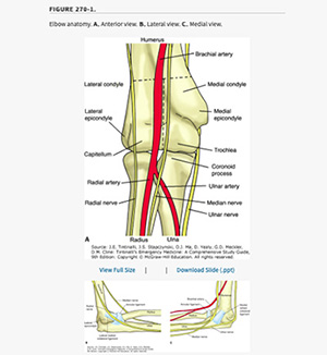
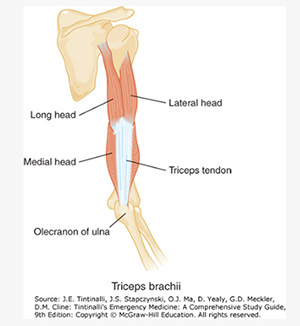
Anatomy
- The triceps muscle is located on the back of the upper arm and has three heads to the proximal tendon, thus the name triceps. The origination points include the infraglenoid tubrical of the scapula and the posterior humerus both superiorly and inferiorly to the radial groove.2 Distally, the triceps tendon inserts onto the olecranon process of the ulna.2 This muscle and its tendons are responsible for elbow extension.
- The muscle is innervated by the radial nerve, which originates from the spinal nerve routes C6 through C8, and runs over the capitellum of the humerus medial to the lateral epicondyle.3
- Nearby nerves include the ulna nerve which runs lateral to the trochlea of the humerus, and the ulnar nerve which runs posteriorly, adjacent to the medial epicondyle of the humerus.4
- Vascularly, the brachial artery runs through the triceps, and provides arterial blood supply to the muscle. The brachial artery then wraps anteriorly around the humerus and branches into the radial and ulnar artery just distal to the anterior elbow joint.5
Injury Presentation:
- History:
- Ruptures of the distal triceps tendon most commonly occur from a fall on an outstretched hand (FOOSH) injury. Due to the extreme eccentric flexion force placed on the triceps muscle, or a direct impact to the insertion point at the olecranon the triceps subsequently detaches from its insertion point.5
- Signs and symptoms of this injury include posterior elbow pain, hearing or feeling a “pop” in the elbow at time of injury, swelling, and ecchymosis.1 5
- The history may also include predisposing factors (see introduction) such a diabetes.
- It’s important to distinguish a triceps tear from an olecranon fracture which also causes a loss of elbow extension. This can be detected with physical exam and x-rays. For this reason xrays are tpyically obtained when evaluating a triceps injury
- Physical exam
- Tears of the distal triceps tendon most commonly occur at the attachment point of the tendon onto the olecranon of the humerus.1
- A sulcus (divot) proximal to olecranon with a more proximal palpable retracted triceps muscle on palpation may be present.5 This would look like a ball of muscle on the back of the arm that does not match the other side.
- Palpation of the distal triceps will illicit tenderness.5
- Strength testing will likely exhibit decreased active elbow extension, however only 20% of cases have complete weakness.1
Diagnostic Testing:
- MRI is the gold standard for diagnosis.1 This imaging can demonstrate soft tissue inflammatory changes of the tendon, as well as the torn and retracted tendon fibers.
- A modified Thompson test can be performed where the elbow is placed with the forearm hanging off a table at a 90-degree angle. The triceps muscle is squeezed and observed for subsequent elbow extension. Lack of elbow extension or diminished movement as compared bilaterally is positive for a distal triceps tendon tear/rupture.5
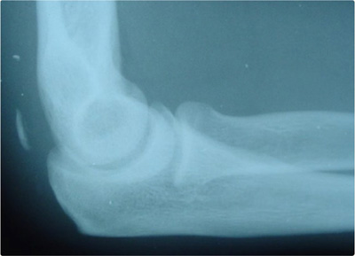
- Lateral elbow x-ray may show avulsed proximal humeral bone if an avulsion injury occurred, known as a ‘fleck’ sign.1 However, if no avulsion is present, the x-ray will appear normal.
- Point of care ultrasound can be used to identify fusiform swelling and retraction of the distal triceps tendon.6
Treatment:
- Initial treatment consists of ice, immobilization in the form of a sling, and analgesics for pain control.5
- Typically the arm feels the best when held in relative extension (straighter), but it’s ok to maintain the arm in the position that is most comfortable.
- Definitive treatment is a surgical repair of the tendon.5 All surgical repair options include reattaching the distal triceps tendon back to its insertion point on the olecranon process.
- In any person who does not have medical reasons that make them unfit for surgery, surgery is typically recommended.
- Surgical treatment options:7
- Transosseous repair: There are two main techniques of repairs.
- The first technique is described by James Paci, MD. In this technique thick suture tied in multiple knots is passed through the triceps in a bridge type pattern running down both the medial and lateral sides of the distal tendon. The suture is then tunneled through the proximal ulna through two drill holes, one medially and one laterally tunneling through the posterior ulna. The suture tails are then anchored into the ulna between the suture exit holes.8 This technique still uses a suture anchor for reinforcement, but requires less of these devices.
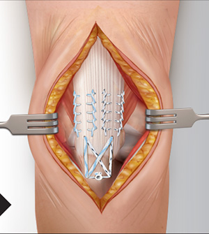
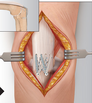
- Suture Anchor Repair
- Suture anchor repairs are typically stronger but require implants (usually non-metal suture anchors) that are placed into the olecranon bone to anchor the tendon back in place.
- The second technique is a variation on the SpeedBridge™ technique commonly used for rotator cuff repairs, described by Paul Caldwell, MD. In this technique two drill holes are made in the proximal olecranon where multiple sutures are anchored into the bone. The sutures are then passed through the distal triceps in a mattress pattern. Two distal olecranon drill sockets are made into which the suture is anchored. This is the preferred technique at Mammoth Hospital as it allows for the repair to lay flat on the patients elbow, reducing the chance for post-operative pain and irritation at the surgical site.8
- Transosseous repair: There are two main techniques of repairs.
- Risks and Benefits of Surgery:
- Risks:
- Bleeding
- Radial nerve injury
- Pain
- Risks:
- Recovery: A full recovery from a triceps tendon repair takes about six months. However, this does not mean there will be complete immobilization without function during this entire period. The elbow will be braced until full elbow extension is achieved, typically over 6 weeks. This is followed by gradual strengthening for an additional 3 – 4.5 months. A gradual return to function will occur in a stepwise fashion directed by a skilled physical therapist. Please find the detailed rehabilitation protocol on the MOI website under patient info -> physical therapy guidelines -> Dr. Brian Gilmer -> Elbow.
Dr. Gilmer’s approach:
I generally prefer the Speedbridge type approach for repairs of all triceps injuries that are greater than 50% that have associated weakness and loss of function. I prefer to repair these tendons earlier, within 2 weeks if possible. If more than 6 weeks have passed since injury a graft (a piece of tissue from a cadaver) may be required in order to bridge any gaps caused by retraction of the tendon. This is often assessed intraoperatively, meaning during surgery, and the best decision is made. In most cases, a graft is not required. Patients interested in more information or a consultation should call the office, contact us via the contact page on the website, or complete the remote consultation form.
References:
- Demirhan M, Ersen A. Distal triceps ruptures. EFORT Open Reviews. 2016;1(6):255-259. doi:10.1302/2058-5241.1.000038
- Triceps Brachii. UW Radiology. Accessed October 28, 2022.
- Chow YC, Lee SW. Elbow and Forearm Injuries. In: Tintinalli JE, Ma OJ, Yealy DM, et al., eds. Tintinalli’s Emergency Medicine: A Comprehensive Study Guide. 9th ed. McGraw-Hill Education; 2020. Accessed October 28, 2022.
- Ulnar Nerve Entrapment at the Elbow (Cubital Tunnel Syndrome) - OrthoInfo - AAOS. Accessed October 28, 2022.
- Elbow and Forearm Injuries | Tintinalli’s Emergency Medicine: A Comprehensive Study Guide, 9e | AccessMedicine | McGraw Hill Medical. Accessed October 28, 2022.
- Tagliafico A, Gandolfo N, Michaud J, Perez MM, Palmieri F, Martinoli C. Ultrasound demonstration of distal triceps tendon tears. Eur J Radiol. 2012;81(6):1207-1210. doi:10.1016/j.ejrad.2011.03.012
- Edelman D, Ilyas AM. Triceps Tendon Anatomic Repair Utilizing the “Suture Bridge” Technique. J Hand Microsurg. 2018;10(3):166-171. doi:10.1055/s-0038-1636729
- Transosseous/SpeedBridgeTM Triceps Repair. :12.